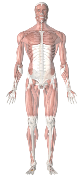
Foot & Ankle

Feet Positioning
Toeing out of the foot is usually associated with lateral hip rotation, which might be due to hypertonic gluteals, deep hip rotators, iliopsoas or adductors. This also implies weakness of medial hip rotators.
Toeing in of the foot is usually associated with medial hip rotation, which might be due to hypertonic tensor fascia latae or anterior fibers of gluteus medius and minimus. This also implies weakness of lateral hip rotators.
Lower Extremity
Feet weight bearing
Begin by looking at the clients shoes. Note the wear pattern on the sole. Is the wear pattern even and symmetrical?
Using a set of scales, have the client place on foot on each scale (checking that the feet are in the same position on each scale). The allowance should only be 1.5kg or 3lbs between the weight recorded on each scale.
Femur angle
Normal Q angle for males is 14 deg and 17 deg for females.Q-Angle is formed in the frontal plane by two line segments
From tibial tubercle to the middle of the patella
From the middle of the patella to the ASIS
Refer to section on Q angle.
Quadriceps muscle tone
Patellae level
Unlevel patellae could indicate structural or functional leg length differential.
Excessive tension in quadriceps could pull a patella in a cephalad direction.
Lumbo- Pelvic Hip Complex
ASIS level
Greater trochanter level
Iliac crest level
Such a pattern often involves an associated series of imbalances characterized by tenderness at the base of the 1st metatarsal, distal medial hamstring attachments, iliolumbar ligament and the superior latissimus dorsi attachment.
Abdominal
Abdominal tonus
Is there an increased degree of tonus in the upper quadrants of the abdomen relative to the lower quadrants?
If so, this may suggest that a faulty respiratory pattern may be present.
Repetitions of sit-ups/curl-ups, slumping postures or ubiquitous forward-leaning postures (such as used by auto mechanics, surgeons, seamstresses, guitarists, manual therapists, etc.) could result in shortening of the upper portions of rectus abdominis and diaphragm.
Is there a visible vertical groove lateral to rectus abdominis?
If so, this suggests predominance of the obliques over the rectus with poor anteroposterior spinal stabilization.
This is to be differentiated from a palpable or visible vertical groove at the mid-line in rectus abdominis (separated linea alba), which could be a result of excessive internal pressure, such as that caused by childbearing.
Distance between the bottom of rib cage and iliac crest symmetrical
Depression of the rib cage might relate to scoliosis and/or to quadratus lumborum, oblique muscles, latissimus dorsi or lumbodorsal fascial shortening, which may, in turn, be due to trigger points within these muscles or in other muscles which refer to these, chronic postural positioning or structural imbalances.
Pelvic elevation may be due to unequal leg length or pelvic obliquity
Torso
Rib and pectoral balance and symmetry
Spinal scoliotic patterns are usually reflected in the positions of the ribs.
Obvious differences in size of comparable muscles can be due to handedness or repetitive use patterns.
Differences in size might also reflect a nerve root lesion at a particular cord level causing atrophy of the apparently smaller muscle.
Differences in rib excursion during breathing could indicate loss of visceral support caudal to the diaphragm, dysfunction of the diaphragm, pleural adhesions or rib restrictions.
Upper Extremity
Arm hang
If not, imbalance in the rotator cuff mechanism and/or an imbalance between flexor and extensor muscle groups associated with the upper crossed syndrome may be present.
Elbows
Are the elbows slightly bent and the tips of the fingers level with each other?
Excessive bend of the elbow could indicate muscular imbalance of the elbow flexors/extensors or the presence of trigger points within those muscles.
When hands hang unevenly, the shoulder girdle may be unbalanced (see previous step regarding acromioclavicular joints).
Hand position
Are the hands slightly pronated with the dorsal surfaces of the hands facing approximately 45° anteriorly?
It needs to be determined whether deviant positions of the hands are due to the position of the forearm (pronation/supination) or the humerus (lateral/medial rotation).
Excessive forearm pronation could be an indication of shortened pronators (teres or quadratus) due to overuse in pronated position, such as often occurs in massage therapy.
The appearance of pronation could represent humeral rotation due to the medial rotators of the shoulder girdle (pectoralis major, latissimus dorsi, teres major and subscapularis, in particular).
Excessive supination warrants examination of the supinator muscle and biceps brachii as well as the lateral rotators of the humerus.
Trigger point activity should be considered in relation to any such hypertonicity or shortening.
Fingers
Excessive curl of the fingers could indicate hypertonic finger flexors, possibly involving trigger point activity in muscles such as infraspinatus, for example.
Arm positioning
Is the distance between the arms and the torso approximately the same on each side?
When excess or inadequate space exists between the arm and the torso, spinal deviations and other more global features should be looked for, such as leg length inequality or pelvic distortions, which would affect the placement of the torso.
Shoulder Complex
Ear level to shoulder height
Cervical distortion due to biomechanical factors may result in such a deviation or a habitual head tilt might relate to visual or auditory imbalances.
An elevated shoulder could be due to postural compensation necessitated by a spinal scoliosis, pelvic distortion, leg length inequality, unilateral loss of the planter arch or other structural deviation.
The apparently lower shoulder could be depressed by shortening or hypertonia involving shoulder muscles, such as latissimus dorsi.
Balance of muscular development of shoulders
If hypertrophy of upper trapezius exists this suggests the possibility of upper crossed syndrome imbalance with consequent inhibition of the lower fixators of the shoulder.
Excessive bulk of the muscles of one shoulder may be due to habits, such as raising the shoulder to hold the phone to the ear.
Elevation of the first rib by hypertonic scaleni muscles could give the appearance of excessive trapezius bulk. This elevation might also impede lymphatic drainage, resulting in a ‘swollen’ appearance of the supraclavicular fossa region.
Acromioclavicular joints level
The appearance of a ‘high shoulder’ could be due to excessive tension or trigger points of the ipsilateral trapezius or levator scapula.
The contralateral shoulder could be lowered by the latissimus dorsi or other shoulder muscles which are shortened or hypertonic.
This differential may also be a result of postural distortion or skeletal abnormality involving the lower extremity, pelvis or torso (scoliosis, fallen arch, pelvic obliquity, etc.).
Head & Cervical Spine
Head tilt
If the head is off-set, pulling to one side or the other, the causes could relate to pelvic base unleveling, loss of planter arch integrity, compensation for spinal deviations or localized suboccipital/cervical/upper thoracic muscular imbalances.
Some degree of tilting may relate to asymmetrical occipital condyles, which is a common and normal occurrence.
Cranial or facial bone dysfunction (for example, involving dental malocclusion or TMJ problems) might lead to compensatory tilting of the head.
Visual or auditory imbalances/dysfunction can lead to an unconscious tendency to tilt or rotate the head.
Head rotation
Fascial symmetry
The distance from:
Hair line to the bridge of the nose
Bridge to tip of the nose
Bottom of nose to tip of chin should be equal.
This is referred to as the rule of thirds.
Alterations of the facial thirds may be indicative of altered growth and development. A long bottom third may be found in the mouth breather.
Earlobes level
If one earlobe is lower than the other, the cause could be cranial distortion (particularly temporal bone) or head tilt.
If one earlobe is lower than the other, are heavy earrings customarily worn, especially in one ear only?
Earlobes flair excessively
If ears flare this might relate to cranial imbalance (involving external rotation of the temporal bones).
Mandibular
Is the any lateral deviation of the mandible?
